
How to Monitor Embryo Development Naturally with AI
- Feb 16
- 6 min read
Choosing the right tools for embryo monitoring can feel overwhelming when you want fast, affordable fertility treatments that match your needs. The move toward AI-driven systems means you benefit from smarter, more precise embryo tracking without extra stress or cost. This guide explains how AI-powered embryo monitoring technologies help you achieve high-quality results at every step, making your journey smoother whether you’re in Europe, Asia, or North America.
Table of Contents
Quick Summary
Key Insight | Explanation |
1. Equipment Calibration is Crucial | Ensure all embryo monitoring tools are calibrated and sterilized to maintain accuracy and reduce contamination risks. |
2. Implement AI Imaging Systems | Use AI-driven imaging technologies for non-invasive monitoring to enhance embryo development assessment. |
3. Optimize Time-Lapse Tracking | Continuous time-lapse monitoring captures detailed embryo development, improving selection accuracy and IVF outcomes. |
4. Embrace Automated Scoring | Utilize machine learning for objective embryo quality assessment, reducing human error and enhancing predictive outcomes. |
5. Confirm Embryo Health Accurately | Non-invasive methods integrated with AI ensure reliable evaluation of embryo viability, maximizing chances of successful pregnancy. |
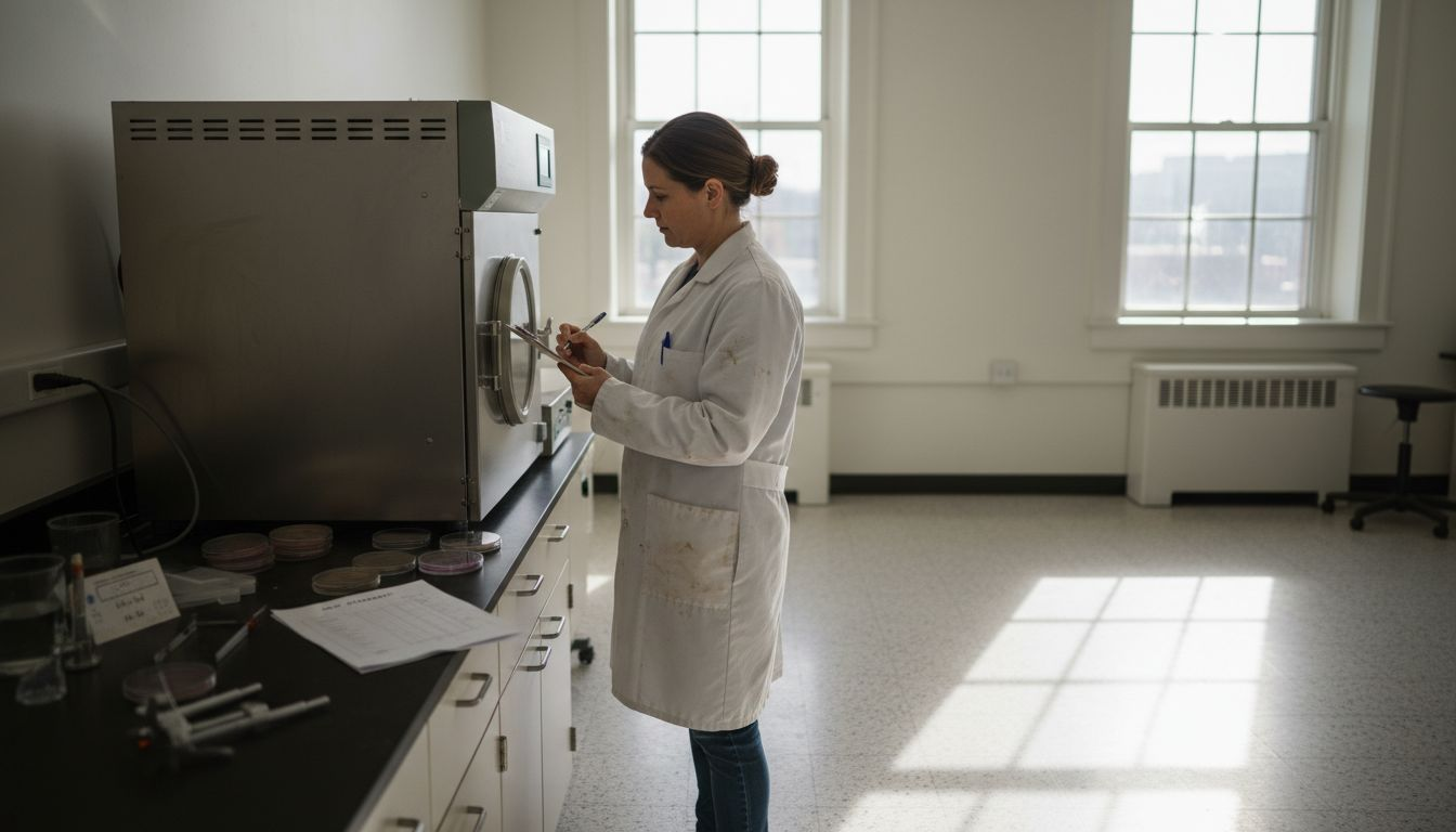
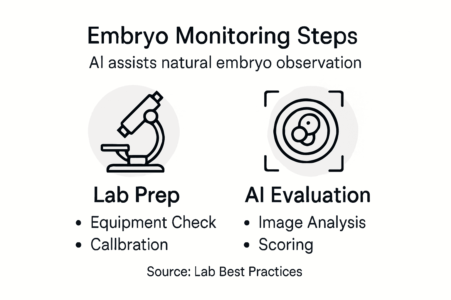
Step 1: Prepare Embryo Monitoring Equipment and Tools
Acquiring and preparing the right embryo monitoring tools is crucial for precise fertility tracking and successful AI-enhanced embryo development assessment. Proper preparation involves selecting specialized equipment that supports accurate observation and data collection throughout the embryonic growth process.
The essential equipment for AI-driven embryo monitoring includes:
Specialized incubators with integrated digital imaging systems
High-resolution microscopes with digital capture capabilities
Advanced robotic preparation systems for consistent culture dish management
Temperature and atmospheric controlled environments
Computer workstations with machine learning software
Sterile laboratory consumables (pipettes, dishes, catheters)
Preparing these tools requires meticulous attention to laboratory standards and quality control protocols. Each piece of equipment must be calibrated, sterilized, and configured according to comprehensive laboratory management guidelines to ensure optimal performance and minimal contamination risk.
Pro tip: Always perform a comprehensive equipment check and system calibration before beginning any embryo monitoring procedure to prevent potential errors and ensure the highest level of accuracy.
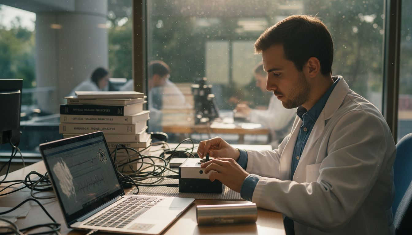
Step 2: Set up AI-driven Imaging Systems for Embryos
Setting up AI-driven imaging systems represents a sophisticated approach to monitoring embryo development with unprecedented precision and minimal invasiveness. These advanced systems combine cutting-edge optical technologies with intelligent machine learning algorithms to capture and analyze embryonic growth in real-time.
The setup process involves several critical components:
High-resolution digital microscopes capable of capturing detailed cellular images
Advanced computer vision algorithms for morphological assessment
Specialized imaging platforms with temperature and atmospheric control
Integrated software with deep learning capabilities
Secure data storage and analysis systems
Implementing these systems requires careful configuration. The label-free optical imaging technology enables non-invasive monitoring, allowing researchers to track embryo development without introducing potentially harmful staining agents or disrupting the delicate cellular environment.
Precision in imaging setup directly correlates with the accuracy of embryo development assessment and potential treatment outcomes.
Pro tip: Regularly calibrate imaging equipment and update AI algorithms to maintain the highest standards of image analysis and embryo monitoring accuracy.
Step 3: Track Embryo Growth Using Time-Lapse Technology
Time-lapse technology represents a groundbreaking approach to monitoring embryo development with unprecedented detail and minimal disruption. By capturing continuous images throughout the incubation process, this advanced method provides researchers with a comprehensive view of critical developmental milestones.
Key components of effective time-lapse embryo tracking include:
Specialized incubation chambers with integrated imaging systems
Convolutional neural network models00264-8/fulltext) for dynamic image analysis
High-frequency image capture mechanisms
Stable environmental control systems
Advanced data processing software
Secure archival systems for developmental sequences
Continuous monitoring technologies enable uninterrupted observation of embryonic growth without removing specimens from optimal culture conditions. This approach allows detailed tracking of morphokinetic changes, providing insights into embryo viability and potential development that traditional intermittent observation methods cannot capture.
Precision in time-lapse imaging can significantly improve embryo selection accuracy and ultimately enhance IVF success rates.
Pro tip: Ensure your time-lapse system maintains consistent temperature and atmospheric conditions to maximize the reliability of developmental tracking.
Step 4: Evaluate Embryo Quality with Automated Scoring
Automated embryo quality scoring represents a revolutionary approach to assessing embryo potential using advanced artificial intelligence technologies. This method transforms subjective visual assessments into precise, data-driven evaluations that significantly improve prediction of implantation success and pregnancy outcomes.
Key components of automated embryo scoring include:
Machine learning algorithms for comprehensive image analysis
Systematic AI-based assessment methods that integrate morphological parameters
Deep neural network classification systems
Real-time developmental stage tracking
Objective morphological feature extraction
Statistical probability calculations for embryo viability
AI integration in IVF laboratories enhances embryo evaluation by analyzing intricate developmental details with unprecedented accuracy. These intelligent systems process multiple parameters simultaneously, reducing human error and providing embryologists with comprehensive, quantitative insights into embryo potential.
Automated scoring transforms embryo assessment from a subjective visual process to a precise, data-driven evaluation method.
Pro tip: Regularly validate automated scoring algorithms against expert embryologist assessments to maintain and improve system accuracy.
Here’s a comparison of traditional embryo assessment versus AI-enhanced approaches:
Aspect | Traditional Assessment | AI-Enhanced Assessment |
Evaluation Type | Visual, subjective | Automated, data-driven |
Consistency of Results | Varies by operator | Highly consistent |
Speed of Analysis | Slower, manual | Rapid, real-time |
Number of Parameters Assessed | Limited, mainly morphology | Multiple, including morphokinetics and genetics |
Error Risk | Higher, human bias | Lower, algorithmic standards |
Data Storage | Minimal, paper-based often | Digital, secure, easily accessible |
Step 5: Verify Embryo Health Before Selection
Verifying embryo health is a critical step in ensuring the highest probability of successful pregnancy. Advanced AI technologies now enable precise, non-invasive assessment of embryo viability, transforming traditional selection methods with unprecedented accuracy and insight.
Key processes for comprehensive embryo health verification include:
Non-invasive imaging techniques
AI-assisted optical health detection without cellular damage
Genetic data integration
Morphokinetic parameter analysis
Cellular nucleus condition assessment
Multi-dimensional risk profiling
Advanced non-invasive evaluation methods combine time-lapse imaging, genetic screening, and machine learning algorithms to create a comprehensive health profile. These intelligent systems analyze multiple biological indicators simultaneously, providing embryologists with a detailed understanding of potential embryo development and implantation success.
Early, accurate embryo health verification significantly increases the likelihood of successful fertility treatments.
Pro tip: Cross-validate AI health assessment results with expert embryologist review to ensure maximum diagnostic accuracy.
The table below summarizes the main methods and benefits at each stage of AI-driven embryo monitoring:
Stage | Key Method | Primary Benefit |
Equipment Preparation | Calibration, Quality Control | Ensures accuracy and reliability |
Imaging System Setup | AI and deep learning integration | Enables non-invasive monitoring |
Time-Lapse Tracking | Automated image sequences | Continuous detailed observation |
Automated Scoring | Machine learning analysis | Objective embryo quality ranking |
Health Verification | Genetic/AI health profiling | Maximizes implantation success |
Enhance Embryo Monitoring with Cutting-Edge AI Solutions
Monitoring embryo development accurately and naturally presents challenges such as maintaining optimal conditions and interpreting complex growth data objectively. This article highlights key pain points like the need for AI-driven imaging systems, time-lapse tracking, and automated scoring to improve embryo quality assessment without invasive methods. If you are seeking a personalized, streamlined approach to fertility care with proven AI technology that addresses these concerns, Aurea Fertility offers just that.
Discover how our clinic leverages advanced AI-assisted protocols to shorten treatment times from months to weeks while enhancing embryo monitoring precision and improving success rates. With features like real-time embryo health verification and AI-driven embryo scoring, we reduce emotional stress and financial burden. Take the next step toward a faster, smarter fertility journey today by visiting Aurea Fertility and learn how our innovative embryo monitoring solutions can support your dreams of parenthood.
Frequently Asked Questions
How can I prepare my lab equipment for AI-driven embryo monitoring?
To prepare your lab equipment for AI-driven embryo monitoring, ensure you have specialized incubators, high-resolution microscopes, and advanced robotic preparation systems. Calibrate and sterilize each piece of equipment according to comprehensive laboratory management guidelines before initiating any monitoring procedures.
What are the benefits of using AI-driven imaging systems for embryo monitoring?
AI-driven imaging systems provide precise, non-invasive observation of embryo development, allowing for real-time analysis without disturbing the fragile cellular environment. Implement these systems to enhance accuracy in capturing detailed cellular images and improve embryo selection outcomes.
How does time-lapse technology improve the tracking of embryo growth?
Time-lapse technology enhances embryo growth tracking by providing continuous, detailed images throughout the incubation process. Use this method to monitor critical developmental milestones without interrupting the optimal culture conditions for the embryos.
What steps are involved in automating embryo quality scoring with AI?
Automating embryo quality scoring involves using machine learning algorithms to analyze images and classify embryos based on morphological parameters. Regularly validate this scoring system against assessments by expert embryologists to ensure high accuracy in evaluating embryo viability.
How can I verify the health of embryos before selection?
To verify embryo health before selection, utilize non-invasive imaging techniques and AI-assisted optical health detection that analyze various biological indicators. Implement this approach to create a comprehensive health profile of each embryo, significantly increasing the chances of successful implantation during fertility treatments.
Recommended
Comments